Phone Number
+61 (03) 9885 4494Email Address
enquiry@melbournevein.com.au
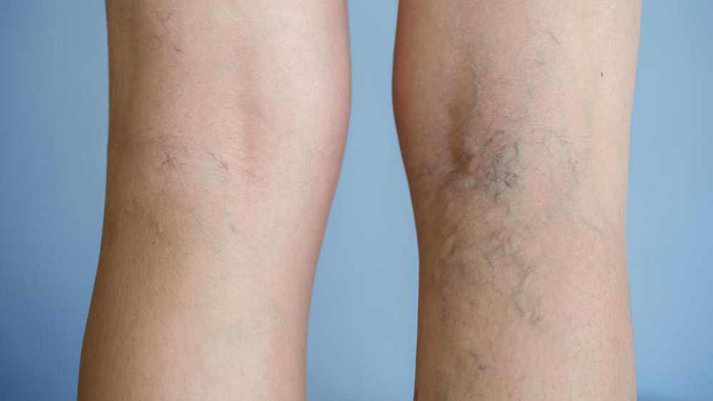
Reticular veins are usually blue or purple in color and may form clusters. They can be associated with pain or discomfort. These are mostly situated in the inner and back of the thighs, back of the legs, ankles, and occasionally on the face. Reticular veins can feed into the spider veins and these feeder veins need to be controlled to successfully treat the smaller veins.
Feeder veins is another name for reticular veins. These Blue Veins look ropy and normally appear on the back of the leg, around the knee. These veins are narrower than Varicose veins but thicker than Spider Veins. It's necessary to treat reticular veins because they're often the veins that feed the networks of smaller spider veins.
All treatments & diagnoses at Melbourne Varicose Vein are performed by Dr. Yazdani.
Reticular veins are extremely common, affecting about 80% of adults, and can be affected by hormonal imbalances, poor veins, or genetic factors. Age, weight, skin damage from UV rays and jobs that include a lot of sitting or standing may all be contributing factors.
In addition, the valves in these reticular veins are ineffective, causing venous blood to flow out toward the skin instead of draining in the usual direction. Since they are deeper and closer to the surface of the skin, they can be more visible. These may cause leg pain, but they may not cause any symptoms at all other than their presence.
These veins have a diameter of around 1-3 mm. They are most often found on the inner thighs or ankles. They can grow on the backs of the legs as well. Reticular veins, unlike varicose veins, do not always protrude above the skin's surface, but they can also be painful or unpleasant. The veins are typically blue-green or purple in color, and they may form uncomfortable clusters.
Treatment techniques for vein problems & lymphatic diseases are very advanced at the Melbourne Varicose Vein clinic. Dr. Nellie is a certified Gold Medal recipient in Sclerotherapy treatment procedures.
| Spider Vein | Reticular Vein | Varicose Vein |
|---|---|---|
| Spider veins are tiny, measuring less than 1 mm | Reticular are a little larger vein measuring 1-3 mm in size | Varicose Veins are larger than 3 mm in size |
| Blue or red in color | Blue-green or purple in color | Dark purple or blue in color |
| Remain beneath the surface of the skin | Usually do not bulge on the skin | Lie under the skin and sometimes bulge or swell out |
| Look like a web structure | These form cluster-like shapes | Bulgy veins, swollen & twisted |
| Found on Lateral thigh & upper 1/3 rd of calf, ankles abdomen chest & face | Seen in the inner & back of thighs, legs ankles & sometimes face | Visible on the thighs, the backs and fronts of the calves, or the inside of the legs near the ankles and feet. |
| These are not painful but might increase due to feeder veins | These can be painful and cause discomfort | These are generally painful and enlarged veins that cause swollen legs & heaviness |
Reticular Veins can be treated by Direct Vision Microsclerotherapy or Ultrasound Guided Sclerotherapy
Dr. Yazdani has achieved great results in restoring vein health.
Call Us or Book an appointment online to visit the Dr. at your convenience.
